
The structure and function of synovial joints is our second dash point under the skeletal system. The skeletal system has a number of different joint types, for example there are fibrous joints and there are cartilaginous joints. You are allowed to ignore this though, as you only need to know about the synovial joints, which are the ones that freely move; such as your hip.
Structure of synovial joints

There are 6 different types of synovial joints:
- Plane (Inter-Tarsal joints) – allows limited gliding movements
- Hinge (Elbow) – allows movements along one axis for flexion or extension
- Pivot (C1 to C2 vertebral joint) – allows rotational movements and some bending
- Ellipsoid/Condyloid (Radius to carpal joint – wrist) – allows movement in two planes, allowing flexion, extension, adduction, abduction, and circumduction.
- Saddle (base of the thumb) – allows same movements as the condyloid but with no axial rotation.
- Ball-and-socket (Hip) – allows movement through three planes (flexion, extension; abduction, adduction; rotation) and is the most mobile of the synovial joints.
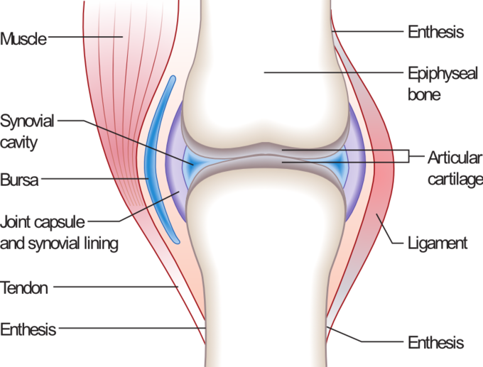
The internal structure of each of these joints, though slightly different in the number of ligaments, tendons, and whether they have menisci or other specialised attributes, is roughly the same. Each joint has:

- Articular cartilage – which helps allow for smooth pain free movement
- A synovial membrane and joint cavity – which hold in the synovial fluid which provides the lubricant for the joint to move smoothly and pain free.

- On top of this layer (usually, though there are some such as the anterior and posterior cruciate ligaments in the knee that are within the joint capsule) there are joint ligaments that hold the 2 bones involved in the joint together and help to restrict movement of that joint to how the synovial joint type moves.
- Although not part of the skeletal system or technically the structure of a joint, muscle tendons are the next layer around joints and may have a bursa (pad for cushioning) between them and the joint to prevent wear and tear. Tendons join muscle to bone and stretch across joints to allow the joint to move.
- Some joints, such as the knee, also have a meniscus in them. This is essentially an extra layer of cartilage that aims to provide cushioning within the joint. It is normally found in joints that bear large loads, such as the knee.
Function of synovial joints
The function of synovial joints first and foremost to provide movement and then to provide stability. Synovial joints are particularly important for movement as they are the type of joint that allows for large movements to happen. Without them, we would have very stiff movements. The structures within the synovial joint that allows them to serve this function are the: synovial fluid, cartilage, and muscles/tendons. These allow for greater movement at these joints than all other joints.
Synovial joints along with other joints of the skeletal system also provide stability. This stability is provided by the joint capsule and ligaments in the joints structure. When the tissues are damaged then the stability of the joint and our body (depending on joint location) is in jeopardy. Furthermore, the type of synovial joint also adds stability to the joint. The ball-and-socket joint is by far the least stable and is the easiest to dislocate, while the plane joints are very stable.
Resources
http://ovrt.nist.gov/projects/vrml/h-anim/jointInfo.html
https://www.boundless.com/biology/textbooks/boundless-biology-textbook/the-musculoskeletal-system-38/joints-and-skeletal-movement-217/classification-of-joints-on-the-basis-of-structure-and-function-820-12063/